
New article by Perrine Chaurand, Wei Liu, Daniel Borschneck, Clément Levard, Mélanie Auffan, Emmanuel Paul, Blanche Collin, Isabelle Kieffer, Sophie Lanone, Jérôme Rose & Jeanne Perrin on "Multi-scale X-ray computed tomography to detect and localize metal-based nanomaterials in lung tissues of in vivo exposed mice"
Abstract :
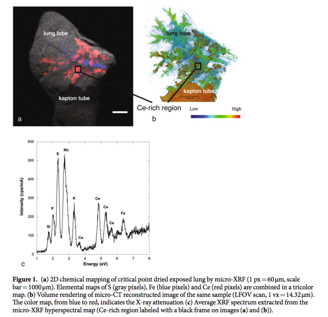
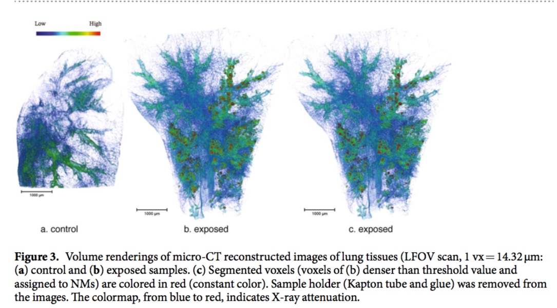
In this methodological study, we demonstrated the relevance of 3D imaging performed at various scales for the ex vivo detection and location of cerium oxide nanomaterials (CeO2-NMs) in mouse lung. X-ray micro-computed tomography (micro-CT) with a voxel size from 14 μm to 1 μm (micro-CT) was combined with X-ray nano-computed tomography with a voxel size of 63 nm (nano-CT). An optimized protocol was proposed to facilitate the sample preparation, to minimize the experimental artifacts and to optimize the contrast of soft tissues exposed to metal-based nanomaterials (NMs). 3D imaging of
the NMs biodistribution in lung tissues was consolidated by combining a vast variety of techniques in
a correlative approach: histological observations, 2D chemical mapping and speciation analysis were performed for an unambiguous detection of NMs. This original methodological approach was developed following a worst-case scenario of exposure, i.e. high dose of exposure with administration via intra- tracheal instillation. Results highlighted both (i) the non-uniform distribution of CeO2-NMs within the entire lung lobe (using large eld-of-view micro-CT) and (ii) the detection of CeO2-NMs down to the individual cell scale, e.g. macrophage scale (using nano-CT with a voxel size of 63 nm).
http://dx.doi.org/10.1038/s41598-018-21862-4